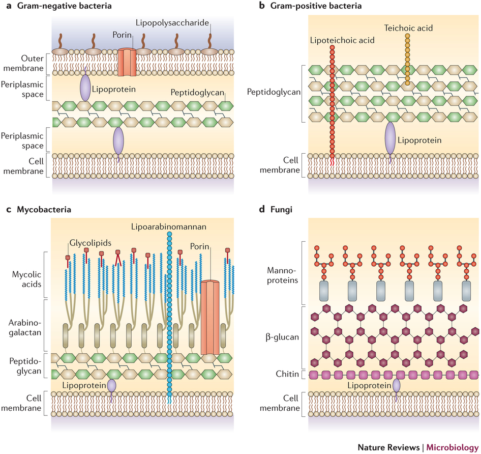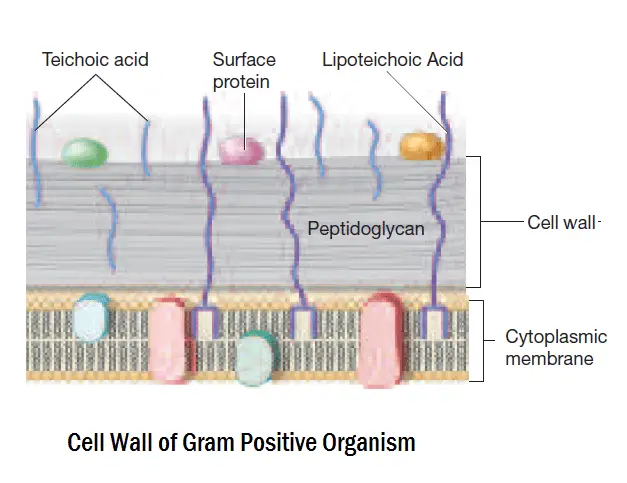

either to the cytoplasmic membrane or cell wall components. Proteins displayed on the cell surface of Gram-positive bacteria must be attached to the cell envelope, i.e. Except for teichoic and teichuronic acids, the structure and biosynthesis of other SCWPs are largely unknown. lipoglycans and lipoteichoic acids ( Fischer, 1994). teichuronic acids or tethered to a lipid anchor moiety, e.g. The SCWPs present in various proportions are either covalently linked to the peptidoglycan backbone, e.g. Besides peptidoglycan, the rigid cell wall of Gram-positive bacteria contains large amounts of wall-associated polymers, also called ‘secondary’ cell wall polymers (SCWPs), which can be classified into three distinct groups: (i) teichuronic acids, (ii) teichoic acids, including lipoteichoic acids and (iii) other neutral or acidic polysaccharides, which cannot be assigned to the two former groups, e.g. polyol phosphate polymers, not found in Gram-negative bacteria ( Shockman & Barrett, 1983). the cytoplasmic membrane and (ii) a cell wall composed at least of peptidoglycan, consisting of linear polysaccharide chains cross-linked by short peptides, covalently linked to teichoic acids, i.e. Therefore, it appears clearly that when employing the term ‘Gram-positive bacteria’ it is extremely important to specify if we refer to a Gram staining result, a cell envelope organization and/or a taxonomic group.įor the purpose of the present review, the term Gram-positive will be used to describe bacteria with a cell envelope composed of (i) a single biological membrane (monodermita), i.e. Even in some deep branches of the phylum Firmicutes, some bacteria clearly exhibit Gram-negative cell envelope ( Shatalkin, 2004). Some other phyla, like BVI Chloroflexi or BVII Thermomicrobia, regroup both monoderm and diderm bacteria ( Botero et al., 2004). Thermotoga maritima in phylum BII Thermotogae ( Snel et al., 2005) or Fusobacterium nucleatum in phylum BXXI Fusobacteria ( Mira et al., 2004). Some bacteria clearly possessing a Gram-negative-like cell envelope architecture are in fact phylogenetically related to the taxonomic group of Gram-positive bacteria, e.g. some members of the phylum BIV Deinococcus- Thermus. More confusingly, some bacteria not taxonomically-related to Gram-positive bacteria retain the Gram staining, e.g. in the genus Mycoplasma or (iii) the presence of a waxy outer sheath preventing penetration of the stain, e.g. in the genus Clostridium, (ii) the absence of a cell wall, e.g. Importantly, some of these Gram-positive bacteria do not retain the Gram stain, either because of (i) a too thin peptidoglycan layer, e.g.

phylum BXIII Firmicutes and those that have high G+C moles percent, i.e.
#GRAM POSITIVE VS GRAM NEGATIVE CELL WALLS MANUAL#
In the Bergey's Manual ( Garrity, 2001), Gram-positive bacteria are divided into those that have low G+C moles percent, i.e. Molecular analyses further revealed that contrary to Gram-negative bacteria, Gram-positive bacteria corresponds to a phylogenetically coherent grouping of prokaryotes within the domain Bacteria ( Woese, 1987). possess a single cell membrane (monoderm prokaryotes) ( Gupta, 1998). two distinct membranes (diderm prokaryotes), Gram-positive have a thick cell wall but lack an outer membrane, i.e.

While the cytoplasmic membrane of Gram-negative bacteria is surrounded by a thin cell wall beneath the outer membrane, i.e. The difference in staining was further related to profound divergence in structural organization of the bacterial cell envelope. retaining or not the stain and thus called Gram-positive and Gram-negative bacteria, respectively. With this method, slightly perfected over the years, bacteria are separated into two groups, i.e. It was in 1884 that the Danish pharmacologist and physician Hans Christian Jaochim Gram published the most-famous staining method in the world of bacteriology ( Gram, 1884). Gram-positive bacteria, cell surface display, membrane protein, cell wall anchoring, S-layer protein, bacterial surface organelle, bacterial protein secretion Introduction


 0 kommentar(er)
